Tech Portfolio

Mathematics

Applied Mathematics in Proton Radiation Therapy

Spot On: Applied Mathematics in Proton Radiation Therapy

Researcher: Prof. Yair Censor

Background

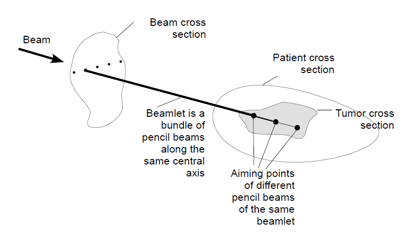
Proton therapy is an advanced form of radiation therapy that uses protons instead of x-rays to destroy cancer cells. Protons can be accelerated to high speeds not far from the speed of light and deposit most of their energy just before they stop, for example, deep in the body, a phenomenon known as Bragg peak (named after William Henry Bragg who discovered it in 1903). This is quite different from x-rays, still most commonly applied in radiation therapy, which deposit an exponentially decreasing amount of energy as a function of tissue penetration depth. As such, with proton therapy one can achieve highest doses at any desired depth, unlike x-ray radiation therapy where the highest dose is from single beams and is always close to the surface. Advanced versions of x-ray and proton therapy utilize many small beams, called beamlets for x-rays and pencil beams for protons. These small beams vary in intensity (they are modulated) to create the dose pattern prescribed by the radiation oncologist, with the purpose of delivering a homogeneous, high dose to the tumor and reduced doses to surrounding normal tissues. This technique, intensity modulated radiation therapy (IMRT) for x-rays and intensity modulated proton therapy (IMPT) for protons, evolved during the 1990s. A mathematical solution of beamlet or pencil beam intensities that leads to acceptable (feasible) dose distribution solves the inverse treatment-planning problem. In 1988, Censor, Altschuler, and Powlis presented the first mathematical algorithm to solve the fully-discretized inverse problem in IMRT. For proton therapy, the same mathematical principles apply, but the solution of an inverse IMPT planning problem is more complex and requires more computer power. Modern proton radiation therapy utilizes actively scanned pencil beams. With the introduction of single-room solutions, it is growing at a rapid pace. Finding efficient mathematical algorithms that can solve the inverse problem in IMRT and IMPT is a very active field of research.


Intensity-Modulated Proton Therapy for Precisely Targeted Cancer Therapy

In modern intensity modulated proton therapy, a large number of actively scanned proton pencil beams reach the tumor and require adjustment of location, depth, and intensity. Finding the right intensity for each of the beams is an intricate mathematical problem and its solution requires advanced mathematical algorithms.
Prof. Yair Censor of the University of Haifa’s Department of Mathematics, in collaboration with  Prof. Reinhard Schulte, a physician scientist at Loma Linda University in California, has developed an innovative mathematical solution method to solve the inverse IMPT planning problem. In practice, the solutions found are most beneficial when one needs to sculpt the dose away from nearby critical organs at risk without compromising the dose to the tumor. This is a common problem when doctors use proton or ion therapy to treat tumors in the brain and the head and neck region.

Research Status

Dr. Censor and his collaborators, experts in computer science and proton therapy, have developed advanced mathematical algorithms to find superior solutions for inverse planning of IMPT and IMRT. One recent development has been to find solutions that allow prescribed under or overdosing of certain dose regions (tumors and organs at risk), specified in clinical trial protocols. Currently they work on methods that deliver solutions that are more homogeneous in dose for individual regions and are more robust to delivery uncertainties such as proton range errors. Another area of recent work is to utilize the benefit of reduced range uncertainty and the possibility to image moving targets with particle imaging techniques for finding better planning solutions for IMPT in challenging cases. In a recent collaboration, with the Heidelberg Ion Therapy (HIT) center we are working on using our algorithms to create carbon ion beams of uniform biological effectiveness using simulated information on the formation of clustered ionization depositions in nanometric, DNA-like cylindrical volumes.

IP Status
US patent granted – Intensity Modulated Proton Therapy (US 9,220,920 B2)

This email address is being protected from spambots. You need JavaScript enabled to view it.


Related pages

Yair Censor, Prof. - Researcher page

Proton Imaging for Improving Proton Radiation Therapy


Proton Imaging for Improving Proton Radiation Therapy

Proton Imaging for Improving Proton Radiation Therapy

Researcher: Prof. Yair Censor


Background

Proton radiation is one of the most advanced forms of radiation therapy to treat cancer. Similar to x-rays, protons release radiation energy to kill cancer cells; but the largest energy release occurs at a depth that is adjustable by the initial proton energy and forms the Bragg peak, formed by all charged particles heavier than electrons (named after William Henry Bragg who discovered it in 1903). In order to assure accuracy in treating a patient with proton Bragg peaks, the medical team must “see” the location of the tumor cells and the path length of protons needed to stop inside the tumor. Thus, one uses imaging for planning the proton treatment as well as for verifying the accurate position of proton Bragg peaks at the time of treatment, which proton therapy experts call pre-treatment “range-verification”. Currently, proton therapy centers use x-ray CT to facilitate treatment planning, but this method is not as accurate as desired (to the millimeter) and often compromised by imaging artifacts as well as substantial additional imaging dose when one uses it frequently during a course of treatment.

Proton radiation therapy has been growing in popularity in recent years, especially if doctors need to deliver very high radiation doses close to critical organs at risk for side effects, which is often the case for brain and head and neck tumors. Further expansion is expected with the possibility to integrate less costly single-room proton equipment into existing radiation therapy centers.

Imaging with Protons - Improving Proton Therapy
Prof. Yair Censor of the University of Haifa’s Department of Mathematics has collaborated for many years with Prof. Reinhard Schulte, a physician scientist at Loma Linda University, and his collaborators in medical physics and computer science, in developing advanced proton imaging technology and fast and effective image reconstruction techniques that run on computer clusters. The same protons that are used for therapy can also be used for imaging, but, remarkably, at about 1000 times lower dose and without the artifacts commonly seen with x-ray CT. The reconstruction algorithms can handle millions of individual protons that are going through the patient and tracked individually to estimate their path and their water-equivalent path length when traversing the patient. This allows reconstruction of the volumetric distribution of the relative stopping power of the tissues, i.e., the property needed for treatment planning and range verification immediately before treatment.

Research Status
Dr. Censor and the US proton CT (pCT) collaboration have developed a preclinical scanner and advanced image reconstruction techniques currently undergoing testing at the Northwestern Medicine Chicago Proton Center (NMCPC). The recent test results have been very promising in that the research team has demonstrated that they can determine proton range with 1 mm or better accuracy with imaging doses that are about 10 times less than with comparable low-dose x-ray CT scans.
A preliminary test performed at the Heidelberg Ion Therapy (HIT) Center also gave good results with helium ions, showing that the helium imaging technology may be suitable for ion radiation therapy.

IP Status
US patent granted – Systems and methodologies for proton computed tomography (US 9,207,193 B2)

This email address is being protected from spambots. You need JavaScript enabled to view it.


Related pages

Yair Censor, Prof. - Researcher page

Spot On: Applied Mathematics in Proton Radiation Therapy


The Language and Numbers Laboratory

The Language and Numbers Laboratory

Welcome to the website of the Language Laboratory. Our lab is affiliated with the Faculty of Education at the University of Haifa and is headed by Dr. Anat Prior. It is part of the Edmond J. Safra Brain Research Center for the Study of Learning Disabilities and the Department of Learning Disabilities.

We investigate high-level human cognitive functions using experimental psychology, educational research, and electrophysiological approaches (Event-Related Potential Technique - ERP).

Our main courses of research:

  • Cognitive consequences of bilingualism
  • Second and foreign language learning
  • Interactions between first and second language systems
  • Minority students' literacy performance
  • Reading comprehension in first and second language.
  • Individual differences in language learning
  • Domain general vs. specific bases of language processing

Our aim is to better characterize and understand the interactions between two languages (or more) in a single neural and cognitive system, and to contribute to the effort to identify the underlying mechanisms leading to individual differences in language learning and processing. Our goal is for this research to foster the development of effective instruction and intervention programs in the domain of foreign language, and programs targeted at minority language students in mainstream education.

carmel logo


Carmel - University of Haifa, Economic Corporation Ltd. 

Eshkol Tower,
25th floor, Room 2502
University of Haifa
Haifa, 3498838
ISRAEL

Tel: +972-(0)4-8288500
Fax: +972-(0)4-8288499
Email: carmel@univ.haifa.ac.il
Website: carmel-ltd.haifa.ac.il

Contact Us

Please contact us for further information

Please type your name.

Please type your last name

Invalid Input

Invalid email address.

Please type your message